Category Archives: sleep

“Insulin Dynamics”

This one has a bit for everyone.


Relationship of Insulin Dynamics to Body Composition and Resting Energy Expenditure Following Weight Loss (Hron et al., 2015)


I think study was actually done a few years ago, originally published here (blogged about here), and re-analyzed through the eyes of Chris Gardner.  I think. (But it doesn’t really matter as the study design appears to be identical.)


Experiment: give someone an oral glucose tolerance test (75 grams glucose) and measure insulin 30 minutes later.  Some people secrete more insulin than others (a marker of insulin resistance); these people also have a lower metabolic rate after weight loss = increased propensity for weight regain.  However, if these people follow a low carbohydrate diet, then the reduction in metabolic rate is attenuated.  Some people who don’t secrete a lot of insulin after a glucose load may do better in the long-run with a lower fat diet.

 

Continue reading

Some nuances of Intermittent Fasting

Intermittent fasting (IF) is not a universal panacea, regardless of whether you’re not eating anything at all for a few days each week/month or just restricting your feeding window to a few hours per day.

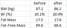
Some protocols, eg, 20h fasting every second day, significantly improve insulin sensitivity in adipose tissue (Halberg et al., 2005). This is expected to make fat gain easier, and while this wasn’t meant to be a study on body composition per se…


body composition


After just a few weeks, things weren’t changing in a good way (NS).


Continue reading

Artificial light regulates fat mass: no bueno.

“despite not eating more or moving less”

We’ve seen this time and time again: LIGHT IS A DRUG.


above quote is extrapolated from this rodent study: “Prolonged daily light exposure increases body fat mass through attenuation of brown adipose tissue activity.”


Artificial light impacts nearly every biological system, and it doesn’t even take very much to have an appreciable effect (think: checking your smart phone or watching a television show on your iPad in bed at night).  In this study, adding 4 hours to the usual 12 hours of light slammed the autonomic nervous system, disrupting sympathetic input into brown adipose leading to a significant increase in body fat  “despite not eating more or moving less.”


Continue reading

LIGHT, Leptin, and Environmental Mismatch

For a long time, the melanocortin system was basically thought to control the color of skin and hair.  It still does, and many redheads are redheaded due to polymorphisms in one of the melanocortin receptors.

Fast forward to 2015: to make a long story short, melanocortins are HUGE players in circadian biology.


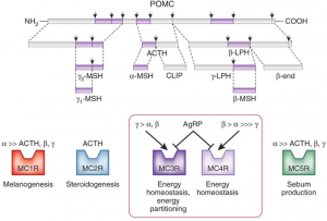
POMC ACTH a-MSH


Brief background (also see figure above):

Fed state -> high leptin -> a-MSH -> MC4R (the receptor for a-MSH) = satiety, energy production, fertility, etc.

Fasted state -> low leptin -> AgRP blocks MC4R = hunger, energy conservation, etc.

MC4R polymorphisms in humans are associated with obesity.  Melanotan II causes skin darkening (marketed as “photoprotection” [no bueno, imo]), enhanced libido, and appetite suppression.


Continue reading

Circadian Disruption Impairs Survival in the Wild

…just read that huge disasters, ranging from Exxon Valdez to Chernobyl, may have been due, in part, to ignorance of basic principles of circadian rhythms.  Gravitas.


circadian rhythms

Continue reading

Entraining Central and Peripheral Circadian Rhythms

“Desynchronization between the central and peripheral clocks by, for instance, altered timing of food intake, can lead to uncoupling of peripheral clocks from the central pacemaker and is, in humans, related to the development of metabolic disorders, including obesity and type 2 diabetes.”

If you haven’t been following along, a few papers came out recently which dissect this aspect of circadian rhythms — setting the central vs. peripheral clocks.

In brief (1):  Central rhythms are set, in part, by a “light-entrainable oscillator (LEO),” located in the brain.  In this case, the zeitgeber is LIGHT.

Peripheral rhythms are controlled both by the brain, and the “food-entrainable oscillator (FEO),” which is reflected in just about every tissue in the body – and is differentially regulated in most tissues. In this case, the zeitgeber is FOOD.

In brief (2):  Bright light in the morning starts the LEO, and one readout is “dim-light melatonin onset (DLMO),” or melatonin secretion in the evening. Note the importance of timing (bright light *in the morning*) – if bright light occurs later in the day, DLMO is blunted: no bueno.

Morning bright light and breakfast (FEO) kickstart peripheral circadian rhythms, and one readout is diurnal regulation of known circadian genes in the periphery.  This happens differently (almost predictably) in different tissues: liver, a tissue which is highly involved in the processing of food, is rapidly entrained by food intake, whereas lung is slower.

Starting the central pacemarker with bright light in the morning but skimping on the peripheral pacemaker by skipping breakfast represents a circadian mismatch: Afternoon Diabetes? Central and peripheral circadian rhythms work together.  Bright light and breakfast in the morning.

Continue reading

Pharmaceutical-grade circadian enhancement?

Is it possible to improve the amplitude and resiliency of your circadian rhythms?  Is this desirable?  Yes and yes, I think.

Go the fuck to sleep.png


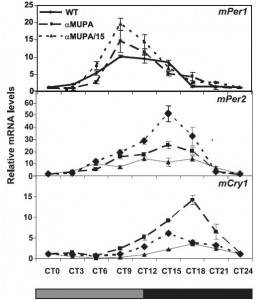
Introducing, the aMUPA mice (Froy et al., 2006).  What you need to know about ’em: they have very robust circadian rhythms.  How is this assessed?  Take some mice acclimated to their normal 12 hour light-dark cycle (LD) and place them in constant darkness (DD).  Then take liver biopsies and measure circadian genes to see how well they still oscillate throughout the dark day; this is also known as the free-running clock, and it craps out differently in different tissues depending on a variety of factors.  Most of the time, however, it’ll run for a few days in the absence of light.  Circadian meal timing also helps to hasten re-entrainment.


Note in the figure below: 1) there are two distinct lines of aMUPA mice; and 2) both exhibit a greater amplitude in circadian oscillations during free-running, or DD conditions.

strong circadian rhythms


Continue reading