Category Archives: liver

Alcohol on keto

This article isn’t about alcohol tolerance.  It’s about your liver.

Tl;dr: with a basic knowledge about alcohol metabolism and ketoadaptation, drinking on keto gives me pause.

It might be nothing, but it gives me pause.

Alcohol is metabolized primarily by alcohol dehydrogenase, producing acetaldehyde and reducing equivalents as NADH.  This pathway produces energy.

Continue reading

AMYLIN

Brief background reading: amylin (according to Wikipedia)


In a study by Hollander on type II diabetics, the synthetic amylin analog pramlintide was tested (Hollander et al., 2003).  In this year-long RCT, over 600 patients were treated with placebo or up to 120 ug pramlintide BID (twice per day).  On average, these subjects were obese (BMI 34), diabetic for ~12 years, and had an HbA1c of 9.1%.  After one year, HbA1c declined 0.62% and they lost about 1.4 kg… not very impressive.


But it’s not all bad news; after viewing those relatively negative results (3 lb weight loss over the course of 1 year), another group of researchers led by Louis Aronne and Christian Weyer believed amylin had yet to be tested proper.  So they designed a better study; it was shorter, used higher doses of pramlintide, and they enrolled obese yet non-diabetic patients (Aronne et al., 2007).  They opted for higher doses of pramlintide (240 ug TID [three times per day]) because in dose-escalation studies, the incidence and severity of adverse drug reactions was consistently low at all doses tested.


They chose to study obese-er subjects (BMI 38, compared to 34 in the Hollander study) because obese subjects lose fat more readily than lean people, so if the study is designed to measure fat loss, then it is better to select a population of subjects where more fat loss is predicted.  They selected non-diabetic subjects for a similar reason; diabetics must regularly inject insulin which promotes the accumulation of fat mass — this could counteract any fat reducing effects of pramlintide.
In other words, it was a more powerful and better designed study.


After 16 weeks, pramlintide-treated subjects lost an average of 3.6 kg (~8 lbs), or about half a pound per week.  30% of patients lost over 15 pounds (1 lb/wk)!  Importantly, the weight loss didn’t appear to have reached a plateau by week 16, so it would have most likely continued along a similar trajectory had the study been longer.  There were no side effects, and a battery of psychological evaluations showed that the patients receiving pramlintide felt it was easier to control their appetite and BW, they didn’t mind the daily injections, and overall well-being increased.  At the very least, these evaluations meant the subjects weren’t losing weight because of nausea or malaise.  In fact, it was quite the opposite.


Continue reading

Tissue-specific fatty acid oxidation

Does it matter where fatty acids are oxidized, liver or skeletal muscle?  Of course, they’re oxidized in both tissues (quantitatively much more in the latter), but relative increases in one or the other show interesting effects on appetite and the regulation of fat mass [in rodents].

Warning: a lot of speculation in this post.

A LOT.

It’s known that LC diets induce a spontaneous decline in appetite in obese insulin resistant patients.  Precisely HOW this happens isn’t exactly known:  the Taubes model?  improved leptin signaling?  probably a little bit of both, other mechanisms, and possibly this one:


Exhibit A. Oxfenicine


oxfenicine

Continue reading

Entraining Central and Peripheral Circadian Rhythms

“Desynchronization between the central and peripheral clocks by, for instance, altered timing of food intake, can lead to uncoupling of peripheral clocks from the central pacemaker and is, in humans, related to the development of metabolic disorders, including obesity and type 2 diabetes.”

If you haven’t been following along, a few papers came out recently which dissect this aspect of circadian rhythms — setting the central vs. peripheral clocks.

In brief (1):  Central rhythms are set, in part, by a “light-entrainable oscillator (LEO),” located in the brain.  In this case, the zeitgeber is LIGHT.

Peripheral rhythms are controlled both by the brain, and the “food-entrainable oscillator (FEO),” which is reflected in just about every tissue in the body – and is differentially regulated in most tissues. In this case, the zeitgeber is FOOD.

In brief (2):  Bright light in the morning starts the LEO, and one readout is “dim-light melatonin onset (DLMO),” or melatonin secretion in the evening. Note the importance of timing (bright light *in the morning*) – if bright light occurs later in the day, DLMO is blunted: no bueno.

Morning bright light and breakfast (FEO) kickstart peripheral circadian rhythms, and one readout is diurnal regulation of known circadian genes in the periphery.  This happens differently (almost predictably) in different tissues: liver, a tissue which is highly involved in the processing of food, is rapidly entrained by food intake, whereas lung is slower.

Starting the central pacemarker with bright light in the morning but skimping on the peripheral pacemaker by skipping breakfast represents a circadian mismatch: Afternoon Diabetes? Central and peripheral circadian rhythms work together.  Bright light and breakfast in the morning.

Continue reading

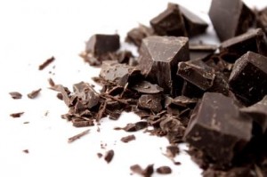
Because chocolate

To improve a memory, consider chocolate –NYT

Dark chocolate could improve memory by 25%, but you’d have to eat 7 bars a day –PBS

Dietary flavanols reverse age-related memory decline –Columbia University Medical Centre

dark chocolate

The actual study: Enhancing dentate gyrus function with dietary flavanols improves cognition in older adults (Brickman et al., 2014)

High flavanol group: 900 mg cocoa flavanols and 138 mg epicatechin (that’d be a LOT of dark chocolate).

Control: 10 mg cocoa flavanols and 2 mg epicatechin

Study duration: 3 months

Funding: NIH & Mars lol

Continue reading

Vegetable oil fatty acids are not essential. 

They are conditionally essential at best, only if docosahexaenoic acid (DHA) is lacking.  We can’t synthesize omega 3 fatty acids, and indeed they do prevent/cure certain manifestations of “essential fatty acid (EFA) deficiency” (Weise et al., 1958), but DHA can do all that and more.  Not that I recommend this, but a diet completely devoid of 18-carbon vege oil fatty acids will not produce EFA deficiency in the presence of DHA. (“vege,” rhymes with “wedge”)

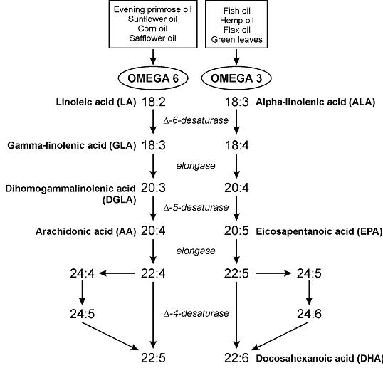
Essential fatty acid metabolism


The “parent essential oils” are linoleic acid (LA) and alpha-linolenic acid (ALA).  The others, which I think are more important and the truly “essential” ones are eicosapentaenoic acid (EPA), arachidonic acid (AA), but mostly just DHA.

The first manifestation of EFA deficiency is dermatitis (Prottey et al., 1975).  Some people say LA is necessary to prevent this, but it would be better phrased as “LA prevents dermatitis;” not “LA is necessary to prevent dermatitis.”  All of the evidence suggesting LA is essential is in the context of DHA deficiency.

Technically, we can convert a bit of ALA to DHA, estrogen helps, testosterone doesn’t (women have better conversion rates)… and I’d speculate that the reverse is probably easier (DHA –> ALA).

Continue reading

Circadian phase delays and metabolism

Remember the “jet lag-resistant” mice?  Guess what: screw with circadian biology and metabolism pays the price.

In brief, vasopressin was classically thought of as an anti-hypotensive hormone.  The vasopressin analog Desmopressin is used to treat bed-wetting.  But vasopressin biology is much more interesting than that: mice lacking both vasopressin receptors require very little time adapting to large circadian phase changes.  And as with many fundamental concepts in chronobiology, this is intimately linked with metabolism.

People with certain polymorphisms of the vasopressin receptor, V1A, exhibit elevated blood glucose levels and are at greater risk for diabetes (Enhorning et al., 2009):

genotype

This risk is strongest in men in the highest quartile of fat intake, and is statistically more significant after adjusting for age and physical activity:

Fat consumption

This study wasn’t designed to be a very powerful indicator of diet-disease relationships, but a little speculation: some think higher fat [and lower carb] intake should be protective against diabetes… which may be true, for people who can tell time.  Alter one nucleotide in the vasopressin 1A receptor gene and the game changes.

Continue reading

Fish, dark chocolate, and red wine.

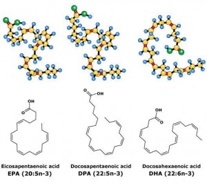
Fish oil fatty acids: EPA & DHA.

I’ve read that EPA tends to show slightly better results in outcomes related to mood, whereas DHA tends to be slightly better for cognition.  Not mutually exclusive; probably a lot of overlap.  This meta-analysis by Martins showed EPA fared better than DHA for depressive symptoms (2009); another one here, stressing the high %EPA relative to %DHA necessary for improvements (Sublette et al., 2011).  Whereas the reverse is true for certain cognitive outcomes in this study by Sinn and colleagues (2012).  Very few studies test EPA vs. DHA directly, and their effects on metabolism are relatively similar.  They’re the ball bearings of fatty acids.epa dpa dha

Continue reading

Going Dutch on Dark Chocolate

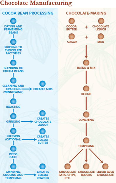
During the production of dark chocolate, cacao beans are fermented, roasted, and processed into 3 components: chocolate liquorcocoa butter, and cocoa powder.  These are combined in various proportions to make unsweetened chocolate.  Sugar can be added to make dark chocolate, or milk & sugar added for milk chocolate.  White chocolate has no cocoa; it’s essentially cocoa butter, sugar, and milk.

ChocolateManufacturingChart

Continue reading

Dietary protein does not negatively impact blood glucose control.

“Dietary protein-derived amino acids have a purpose, and that purpose is not carbs.”

At a reasonable level of dietary intake, protein is used for the maintenance of organs & tissues.  Lean body mass.  It’s functional.  Protein isn’t stored in any appreciable capacity, and most excess is either oxidized or stored as glycogen.  Theoretically, about 50-60% of protein-derived amino acids can be converted into glucose, mathematically, but it’s not what you think…

“At a reasonable level of dietary intake.”  A recent publication took a look at this (Fromentin et al., 2013).   They set out to determine how much protein is converted to glucose under “optimal gluconeogenic conditions.”  That is, the subjects were 12 hours fasted, which is a physiologically relevant, optimal gluconeogenic condition.  They were then given 4 eggs (~23 g protein) that were labeled with two stable isotopes (15N & 13C, derived from hens fed isotope-enriched diets!).  Throughout the entire study duration, the subjects were infused with a third isotope, 2H-glucose.  By collecting and analyzing the enrichment of isotopically-labeled metabolites like expired CO2, urea, and glucose, the researchers were able to determine the fate of those 23 grams of protein.

Some of the dietary protein-derived amino acids were used for protein synthesis, others were oxidized.  But blood glucose levels did not change.  Nor did the rate of gluconeogenesis.

Continue reading